Header
Sir Ganga Ram Hospital, New Delhi

Liquid Biopsy for Cancer: How It Works and Why It Matters

Liquid biopsy for cancer: What it is, how it works, and why it matters in India

A blood test that’s changing how cancer is detected, monitored, and treated

This page is for informational purposes only and does not replace medical advice. Please consult Dr. Aditya Sarin or a qualified oncologist to understand whether liquid biopsy testing is appropriate for your specific case. As of 2026, this reflects current clinical guidance.

For decades, confirming and characterizing a cancer meant a needle, an endoscope, or surgery, whatever it took to get a physical piece of the tumor. Liquid biopsy changes that equation for many patients, at least in part, by pulling the same kind of genetic information out of a simple blood draw. It isn’t a replacement for everything a tissue biopsy does, but it’s become a genuinely useful tool in several specific situations.

What is a liquid biopsy?

According to the National Cancer Institute, a liquid biopsy analyzes bits of tumor material, mainly DNA, but also RNA, proteins, or whole cells, found in bodily fluids such as blood. The most widely used version looks for circulating tumor DNA, or ctDNA, tiny fragments of DNA that tumor cells shed into the bloodstream as part of their normal life cycle.

This is different from a tissue biopsy, which requires physically removing a piece of the tumor, sometimes through a large needle, an endoscope, or open surgery. According to MD Anderson Cancer Center, ctDNA fragments are distinguishable from ordinary cell-free DNA released by healthy, dying cells, mainly by their shorter length, which is part of what makes the test technically possible.

How liquid biopsy testing works

Once a blood sample is drawn, laboratory technology analyzes it for the presence and quantity of ctDNA, along with any specific genetic mutations it carries. Because tumors constantly release DNA as their cells divide and die, a growing tumor generally sheds more ctDNA, and a shrinking one sheds less, which is part of what allows the test to track a cancer’s behavior over time rather than just confirm its presence once.

There are two broad approaches. Testing for specific known mutations is useful once a cancer type is established and doctors want to check for one particular genetic change, while broader genomic profiling screens ctDNA more comprehensively across many genes at once, often used when the exact treatment-relevant mutation isn’t yet known.

Liquid biopsy vs tissue biopsy: What each one actually tells you

Tissue biopsy remains what the National Cancer Institute calls the gold standard for diagnosing cancer and examining its cells directly under a microscope, and it’s still generally required to confirm a cancer diagnosis in the first place. Liquid biopsy is better suited to different jobs: identifying mutations that guide targeted drug selection, tracking whether ctDNA levels are falling or rising during treatment, and in some cases picking up early signs that a cancer is coming back, sometimes months before it would be visible on a scan.

The two aren’t interchangeable, and a negative liquid biopsy result doesn’t rule out cancer the way a negative tissue biopsy might. If a liquid biopsy doesn’t detect a mutation that a tissue biopsy would confirm, regulators, including the FDA, have recommended falling back on tissue testing rather than treating the blood test result as final.

Why non-invasive cancer testing matters

The appeal of liquid biopsy is fairly intuitive once you consider what tissue biopsies actually involve. A blood draw is safe, inexpensive, and easy to repeat, while a tissue biopsy carries real procedural risk and generally isn’t something a patient can undergo every few weeks to check on treatment progress. This matters especially for monitoring: since ctDNA can be tracked repeatedly over the course of treatment, it offers a way to watch how a cancer is responding without the burden of repeated invasive procedures.

That said, the technology still has real, acknowledged limitations. According to a 2024 update from the National Cancer Institute, one of liquid biopsy’s biggest technical challenges is simply that tumors, particularly small or shrinking ones, often don’t release enough ctDNA into the bloodstream for current tests to reliably detect. Researchers are actively working to improve this sensitivity, but for now, a negative liquid biopsy doesn’t always mean there’s nothing to find.

Why liquid biopsy matters in India

Precision oncology in India increasingly depends on identifying specific genetic mutations to match patients with targeted therapies, something we’ve covered across several cancer types on this site, from EGFR and ALK testing in lung cancer to BRCA testing in breast and ovarian cancer. Liquid biopsy offers a way to get at this information without always requiring a fresh tissue sample, which matters in situations where re-biopsying a tumor is difficult, risky, or simply not something a patient wants to go through again.

Access to this technology in India is expanding through major cancer centers, though it remains a specialized test used selectively rather than a routine part of every cancer workup. Whether it’s the right tool for a specific situation depends on the cancer type, the question being asked, mutation status, and whether it’s for detection or monitoring, and should be discussed directly with an oncologist.

Note. Recommend confirming with Dr. Sarin which specific liquid biopsy panels or indications his practice currently uses, so the page reflects his actual approach accurately.

The takeaway

Liquid biopsy is a genuinely useful addition to cancer care, particularly for guiding targeted therapy and monitoring treatment response, but it works alongside tissue biopsy and imaging rather than replacing them outright. Understanding what the test can and can’t tell you helps set the right expectations if your doctor recommends one.

If you have questions about whether liquid biopsy testing is relevant to your diagnosis or treatment plan, book a consultation with Dr Aditya Sarin at Sir Ganga Ram Hospital, New Delhi.

References

  1. National Cancer Institute: Liquid Biopsy: Using Tumor DNA in Blood to Aid Cancer Care.
  2. MD Anderson Cancer Centre: Liquid Biopsies: Understanding ctDNA and Circulating Tumor Cells.
  3. National Cancer Institute: Pump Up the Volume: “Priming Agents” May Improve Cancer Liquid Biopsies.

Immunotherapy Side Effects: What Patients Should Know

Immunotherapy Side Effects: What Patients Need to Know Before Starting Treatment

Why immunotherapy side effects look different from chemo and what to watch for 

This page is for informational purposes only and does not replace medical advice. Please consult Dr Aditya Sarin or a qualified oncologist about your specific treatment plan and what side effects to expect. As of 2026, this reflects current clinical guidance.

If you’re about to start immunotherapy, you’ve probably already heard it described as “gentler” than chemotherapy. In many ways, that’s fair; many patients tolerate it well. But immunotherapy side effects work through a completely different mechanism than chemo side effects, and that difference matters. They can show up later than you’d expect, in places you wouldn’t necessarily connect to a cancer drug, and they need to be reported quickly rather than waited out. Here’s what’s actually going on in your body, what’s common, and what needs urgent attention.

Why immunotherapy side effects are different from chemotherapy

Chemotherapy damages fast-growing cells throughout the body, which is why it causes things like hair loss and nausea. Immunotherapy works through an entirely different mechanism. According to the National Cancer Institute, immunotherapy helps your immune system detect and destroy cancer cells more effectively, and many side effects happen because that revved-up immune system also attacks healthy cells and tissue. In other words, the same process that helps immunotherapy fight cancer also causes side effects, which is part of why oncologists can’t simply turn the treatment “down” the way they might adjust a chemo dose.

This immune overactivation is why immunotherapy side effects are officially called immune-related adverse events (irAEs) rather than just “side effects.” They resemble autoimmune conditions because, functionally, that’s close to what’s happening: your immune system, no longer held in check, starts treating parts of your own body as a threat.

Common early side effects

According to the American Cancer Society, the most frequently reported immunotherapy side effects include:

  • Fatigue
  • Skin changes, including rash or itching
  • Flu-like symptoms such as fever, chills, and muscle aches
  • Stomach issues like nausea, vomiting, or diarrhoea
  • Soreness, redness, or swelling at the injection or infusion site

Most of these are mild and show up early in treatment. They’re also, importantly, not a sign the treatment isn’t working, and they’re not automatically a sign it is either. Report them to your care team regardless, since the same symptom can range from a minor nuisance to the first sign of something that needs medication.

Immune-related adverse events: What to know about the more serious ones

Beyond the common early symptoms, immunotherapy can affect specific organs in ways that need closer monitoring. Per guidance summarised by the National Comprehensive Cancer Network and echoed across major cancer centres, the organs most commonly affected include:

  • Lungs (pneumonitis): New or worsening cough, shortness of breath, chest pain
  • Gut (colitis): Diarrhoea, blood in stool, severe abdominal pain
  • Liver (hepatitis): Yellowing of the skin or eyes, dark urine, right-sided abdominal pain, unusual fatigue
  • Endocrine glands (thyroiditis, adrenal insufficiency): Unexplained weight change, feeling unusually cold or hot, extreme tiredness, dizziness
  • Skin: Rash, blistering, painful sores

Thyroid problems are worth knowing about specifically. Immunotherapy-related thyroid damage is often permanent once it happens, meaning patients may need thyroid hormone replacement medication for life, according to oncology nurse practitioners who monitor these effects closely. It’s manageable, but it doesn’t reverse on its own once treatment ends.

Most immune-related adverse events, according to the Cancer Council, tend to appear several weeks or months after starting treatment rather than immediately, and in rare cases can appear even after treatment has finished. That delay is part of why ongoing monitoring matters throughout, not just at the start.

Immunotherapy and fatigue

Fatigue shows up with immunotherapy for different reasons than it does with chemo. It’s not usually tied to anaemia or bone marrow suppression the way chemo fatigue often is. Instead, it can be a general immune-system response, or, in some cases, an early sign of an endocrine problem like thyroid or adrenal involvement, both of which commonly cause tiredness.

That distinction matters practically. If fatigue during immunotherapy is new, worsening, or paired with other changes, weight shifts, feeling unusually cold, or dizziness, it’s worth flagging to your oncologist rather than assuming it’s simply treatment-related tiredness. A quick blood test can usually tell the difference.

When immunotherapy side effects need urgent attention

Immune-related adverse events can escalate faster than people expect, which is why prompt reporting matters more here than with most cancer treatments. Symptoms that need a same-day call to your oncology team, or emergency care if you can’t reach them, include:

  • Severe or worsening shortness of breath, or new chest pain.
  • Severe diarrhoea, or blood in your stool.
  • Yellowing of the skin or eyes.
  • Confusion, severe headache, neck stiffness, or vision changes.
  • Severe muscle weakness or new numbness.
  • A fever that doesn’t settle, especially alongside any of the above.

Even mild versions of these symptoms are worth mentioning early. Immune-related adverse events are far easier to manage when caught before they progress, and most are reversible with prompt treatment, typically corticosteroids to calm the immune response, when addressed quickly.

How your medical oncologist manages immunotherapy side effects

Because irAEs can appear late and affect almost any organ, monitoring during immunotherapy is often more structured than during chemo. A medical oncologist like Dr Aditya Sarin typically orders regular bloodwork to catch early thyroid, liver, or kidney changes before symptoms appear, asks about new or unusual symptoms at every visit, and has a clear plan for grading severity and stepping in with steroids or other treatment when something crosses the line from mild to concerning. This kind of active surveillance is part of what makes immunotherapy safer in practice than it might sound on paper.

The takeaway

Immunotherapy side effects work differently from chemo side effects, and understanding that difference, especially the delayed timing and the wide range of organs that can be affected, helps you know what’s worth reporting and when. If you’re considering immunotherapy or already partway through treatment, book a consultation with Dr Aditya Sarin to talk through what to expect and how your specific treatment plan will be monitored.

References

Chemotherapy Side Effects: What to Expect and Manage

Chemotherapy Side Effects: What to Expect and How to Manage Them

What actually happens to your body during chemo, and the practical steps that help

This page is for informational purposes only and does not replace medical advice. Please consult Dr Aditya Sarin or a qualified oncologist about your specific treatment plan and what side effects to expect. As of 2026, this reflects current clinical guidance.

If you’ve just started chemotherapy, or you’re about to, the side effects list can feel like the scariest part of the whole process. Some of that fear is fair. Chemo does affect the body beyond the tumour it’s targeting. But a lot of what people picture- constant vomiting, total hair loss for everyone, being wiped out every single day- isn’t how it plays out for most patients anymore. Anti-nausea medication in particular has come a long way over the past couple of decades. Here’s what’s actually common, why it happens, and what helps.

Why chemotherapy causes side effects in the first place

Chemotherapy drugs work by attacking cells that divide quickly, which is exactly what cancer cells do. The problem is that several types of healthy cells also divide fast, including cells in the bone marrow, the lining of the mouth and gut, and hair follicles. When chemo affects those cells too, you get the side effects most people associate with treatment. According to the American Cancer Society, your likelihood of experiencing a given side effect also depends on the dose and combination of drugs used, which is why two patients on similar-sounding regimens can have very different experiences.

Fatigue

Fatigue is widely reported as the single most common side effect of chemotherapy, and it isn’t the same as normal tiredness. It can be deep and persistent, largely unrelated to how much sleep you got, and it tends to peak a few days after each treatment session before easing off until the next one. The American Cancer Society’s guide to cancer-related fatigue notes that fatigue can come from the cancer itself, the treatment, or both, and that it’s worth reporting to your care team rather than assuming it’s something you just have to push through.

A few things that genuinely help:

  • Plan demanding tasks for the days you know you tend to have more energy, rather than fighting through low days
  • Short naps during the day, alongside normal sleep at night, rather than one or the other
  • Light, regular movement. It sounds counterintuitive when you’re exhausted, but gentle exercise, even a short walk, tends to reduce fatigue rather than worsen it
  • Let people actually help with errands and chores instead of pushing through alone

If fatigue is affecting your ability to function day to day, or it’s not easing at all between sessions, tell your oncologist. Sometimes fatigue has a treatable cause underneath it, like anaemia, rather than being chemo itself.

Nausea and vomiting during chemotherapy

Nausea is probably the side effect people dread most, and historically it was one of the hardest to control. That’s changed a lot. The National Cancer Institute explains that chemotherapy-induced nausea and vomiting can be acute (within 24 hours of treatment), delayed (one to seven days after), or anticipatory (before treatment even starts, often after a rough earlier session), and that anti-nausea medicines given before treatment work well to prevent or reduce it in most patients.

Alongside medication:

  • Small, light meals through the day instead of a few large ones
  • Skip greasy, spicy, or strongly scented food, since smell can trigger nausea even more than taste
  • Keep fluids up, since nausea and vomiting can dehydrate you quickly
  • Sitting upright for a while after eating, rather than lying down, is one of the tips the Mayo Clinic recommends alongside medication

Keep your oncology team updated on how well your anti-nausea plan is actually working. It’s adjustable, and you shouldn’t feel like you just have to get through it.

Hair loss from chemotherapy

Hair loss is one of the most visible and, for a lot of people, emotionally difficult side effects. It happens because chemo drugs damage the fast-dividing cells in hair follicles, and it can affect hair anywhere on the body, not just the scalp. A 2023 review in PMC puts the rate of chemotherapy-induced hair loss at around 65% of patients, though this varies a lot by drug and dose, and not everyone going through chemo loses their hair.

The good news is that it’s temporary for most patients. According to the American Cancer Society, whether and how much hair loss happens depends on the specific treatment, and hair typically starts growing back within a few months of finishing chemotherapy, sometimes with a different texture or colour at first.

Scalp cooling is currently the only FDA-cleared option for reducing chemotherapy-induced hair loss, using a cold cap during treatment sessions to reduce blood flow to the scalp. It doesn’t work for everyone and isn’t suitable for every cancer type, so ask your oncologist whether it’s an option for your specific treatment. Others plan ahead with wigs, scarves, or a shorter haircut before treatment starts, which some people find makes the transition feel less sudden.

Other side effects worth knowing about

Fatigue, nausea, and hair loss get most of the attention, but chemo can also affect:

  • Appetite and taste: food may taste metallic or just unappealing for a while
  • Mouth sores and dry mouth
  • Digestive changes, including diarrhoea or constipation depending on the drug
  • Numbness or tingling in hands and feet (peripheral neuropathy)
  • Lower blood counts, which raise infection risk and can cause easy bruising or bleeding

Not everyone gets all of these, and your oncologist will usually tell you upfront which ones are more likely with your specific regimen.

When a side effect needs urgent attention

Most chemo side effects are unpleasant but manageable at home with your care team’s guidance. A fever is different. Because chemotherapy can lower your white blood cell count, a condition called neutropenia, your body’s ability to fight infection drops too, sometimes to the point where fever is the only warning sign something is wrong. The National Cancer Institute and the CDC both advise calling your care team right away for a temperature of 100.4°F (38°C) or higher, even if you otherwise feel okay, and note that this risk is usually highest 7 to 12 days after a chemo dose, when blood counts hit their lowest point.

Other symptoms that warrant a same-day call include severe or persistent vomiting, chest pain, shortness of breath, unusual bleeding or bruising, or any sign of infection around a catheter or port site.

How your medical oncologist manages side effects through treatment

Side effect management isn’t a separate add-on to chemotherapy; it’s built into the plan from the start. A medical oncologist like Dr Aditya Sarin adjusts anti-nausea protocols, monitors blood counts between cycles, and can modify dosing or timing if side effects become too severe, all while keeping the treatment as effective as possible against the cancer itself. This is part of why regular check-ins during chemo matter beyond just tracking the tumour. Side effects you mention casually can change what happens at your next session.

The takeaway

Chemotherapy side effects are real, but they’re also largely predictable and manageable with the right support. If you’re starting treatment soon or already partway through and something doesn’t feel manageable, book a consultation with Dr Aditya Sarin to talk through what to expect and how to adjust your plan.

References

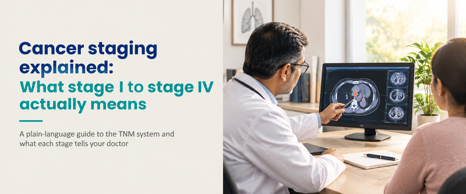
Cancer Staging Explained: Stage I to Stage IV Meaning

Cancer staging explained: What stage I to stage IV actually means

A plain-language guide to the TNM system and what each stage tells your doctor

This page is for informational purposes only and does not replace medical advice. Please consult Dr Aditya Sarin or a qualified oncologist to understand what your specific stage means for your treatment. As of 2026, this reflects current clinical guidance.

Getting a stage attached to a cancer diagnosis can feel like getting a verdict, but staging is really a description, not a sentence. It tells your doctor how much cancer is present and where, which shapes everything from which treatments make sense to how closely you’ll be monitored afterwards. Here’s what the numbers and letters actually mean.

What is cancer staging and why does it matter

According to the American Cancer Society, staging is the process of finding out how much cancer is in a person’s body and where it’s located. Doctors use imaging tests, biopsies, blood tests, and sometimes findings from surgery to determine this.

Staging matters for two main reasons. It helps determine the most appropriate treatment, since an early-stage, localized cancer might be treated effectively with surgery or radiation alone, while a more advanced stage often needs treatment that reaches the whole body, like chemotherapy or targeted therapy. It also gives doctors a general way to estimate prognosis and communicate clearly about a case, both with each other and with the patient.

Not every cancer is staged the same way. Leukemias, for instance, are usually described as untreated, in remission, or recurrent rather than given a numbered stage, since they typically involve the blood throughout the body from the start.

The TNM staging system explained

The most widely used staging system is called TNM, maintained by the American Joint Committee on Cancer. It breaks a cancer down into three components.

T, for tumor, describes the size of the original tumor and how far it has grown into nearby tissue. N, for node, describes whether the cancer has spread to nearby lymph nodes, and how many. M, for metastasis, describes whether the cancer has spread to distant parts of the body.

Each of these gets its own number or letter, for example T2, N1, M0, and the specific combination is then translated into an overall stage, usually written as a Roman numeral from I to IV. Cervical and other reproductive cancers use a closely related system called FIGO staging, developed by the International Federation of Gynaecology and Obstetrics, which maps onto TNM stages fairly directly.

What stage 0 to stage IV actually means

While the exact criteria differ by cancer type, the general pattern across most solid tumor cancers looks like this.

Stage

General meaning

Stage 0

Abnormal cells are present only in the layer where they started, sometimes called carcinoma in situ or pre-cancer. Not every cancer type has a stage 0.

Stage I

A small tumor confined to where it started, with no or minimal spread to lymph nodes.

Stage II

A larger tumor, or limited spread to nearby lymph nodes, still without distant spread.

Stage III

Further local growth or more extensive lymph node involvement, but not yet spread to distant organs.

Stage IV

Cancer has spread to distant parts of the body, also called metastatic cancer.

Stages are sometimes subdivided further with letters, for example stage IIIA versus stage IIIB, to capture finer distinctions within a stage. For cancer-specific examples of how this plays out, breast cancer staging, ovarian cancer staging, and cervical cancer staging each show how the general TNM framework gets adapted to a specific cancer type.

What does stage 4 cancer mean

Stage IV specifically means the cancer has metastasised, spread from where it started to distant organs or tissues elsewhere in the body. This is generally the most advanced stage across cancer types.

Stage IV doesn’t mean the same thing for every cancer, and it doesn’t automatically mean treatment stops being worthwhile. Depending on the cancer type, where it has spread, and how it responds to treatment, some stage IV cancers are managed successfully as a long-term, chronic condition for years, particularly as targeted therapy and immunotherapy options have expanded significantly in recent years.

How staging affects cancer prognosis

This is the part worth being precise about, since staging is most easily misunderstood here. Survival statistics, like a “5-year survival rate,” are always tied to the stage at the time of diagnosis, not to any later restaging. According to the American Cancer Society, if a cancer is diagnosed at stage II and later spreads, the original 5-year survival figures for stage II still apply to that diagnosis; they don’t get replaced by stage IV statistics.

Just as importantly, survival rates differ enormously by cancer type at the same stage. Stage I of one cancer might carry a very different outlook than stage I of another, since staging measures the extent of spread, not how aggressively a particular cancer behaves. Age, overall health, whether the cancer cells carry specific genetic or biomarker changes, and how well the cancer responds to treatment all factor into an individual’s actual prognosis alongside the stage itself.

This is exactly why a stage number on its own, without the specific cancer type and an oncologist’s full context, isn’t something to draw firm conclusions from.

The takeaway

Cancer staging is a tool for planning treatment and understanding the extent of a diagnosis, not a fixed prediction of what will happen. The same stage can carry very different implications depending on the cancer type, which is why the number itself matters less than a full conversation with your oncologist about what it means for your specific situation.

If you’ve received a staging result and want it explained clearly, book a consultation with Dr Aditya Sarin at Sir Ganga Ram Hospital, New Delhi.

References

  1. American Cancer Society. Cancer Staging.

Warning Signs of Cancer Every Indian Should Know

Warning signs of cancer every Indian should know

The changes in your body worth paying attention to, and when to act on them

This page is for informational purposes only and does not replace medical advice. Please consult Dr Aditya Sarin or a qualified oncologist to evaluate any symptoms specific to you. As of 2026, this reflects current clinical guidance.

Cancer doesn’t always announce itself clearly. Some of its earliest signs look exactly like something ordinary: a stubborn cough, a bit of fatigue, indigestion that won’t quit. What matters less is any single symptom; what matters more is persistence. A change that lasts for weeks, gets worse, or has no obvious explanation is worth bringing to a doctor.

Common warning signs of cancer everyone should know

According to the American Cancer Society, some of the most common signs and symptoms that may point to cancer include fatigue that doesn’t improve with rest, unexplained weight loss or gain of 10 pounds or more, swelling or lumps anywhere in the body, and pain that is new, persistent, or has no clear cause.

A few others are worth knowing specifically. A cough or hoarseness that doesn’t go away. Unusual bleeding or bruising with no obvious reason. A change in bowel habits, such as diarrhoea or constipation that lingers, or blood in the stool. Bladder changes, including pain when urinating or blood in the urine. A sore that doesn’t heal, or a mole that changes in size, shape, or colour. Fever or night sweats that show up without an infection to explain them.

Most of these symptoms are far more often caused by something other than cancer, an infection, anaemia, or a benign condition. But that’s exactly why seeing a doctor matters even when cancer feels unlikely, since the underlying cause still needs treating either way.

Cancer symptoms in women

Some symptoms deserve particular attention in women, since they line up with the cancers most common among Indian women, breast, cervical, and ovarian cancer among them. A new lump or thickening in the breast, changes in breast shape or skin, or nipple discharge are classic early signs of breast cancer, which remains the most common cancer among Indian women. Abnormal vaginal bleeding, especially between periods or after menopause, along with unusual discharge or pelvic pain, can point to cervical or uterine cancer. Persistent bloating, pelvic pain, or feeling full quickly, if it lasts more than two to three weeks, is worth checking for ovarian cancer, since this cancer rarely produces clear symptoms until it has progressed.

Cancer symptoms in men

A few symptoms are worth flagging specifically for men. Difficulty urinating, a weak urine stream, or blood in the urine can be linked to prostate or bladder cancer. Never brush off a lump, swelling, or heaviness in the testicles, since testicular cancer often shows up this way and responds well to early treatment. A persistent cough, hoarseness, or coughing up blood are signs worth taking seriously for lung and throat cancers, both of which rank among the leading cancers in Indian men, alongside cancers of the mouth and tongue linked closely to tobacco use.

When to see an oncologist

You don’t necessarily need to see an oncologist as soon as a symptom appears. A general physician is usually the right first stop, and they’ll refer you onward if something needs specialist evaluation. That said, a few situations call for moving faster: a symptom that has lasted more than two to three weeks without an obvious cause, a lump that doesn’t go away, unexplained weight loss, or any bleeding that isn’t normal for you. If you already have a strong family history of a particular cancer, it’s also reasonable to ask your doctor directly whether an oncology consultation makes sense sooner rather than later.

An oncologist’s evaluation typically starts with your history and a physical exam, followed by imaging or blood tests as needed, and a biopsy only if something specific needs confirming

. Getting this sequence started early rarely costs you anything except peace of mind, while delaying it can cost real time if something does turn out to be cancer.

Cancer in India, why awareness matters

According to estimates published in the Indian Journal of Medical Research based on National Cancer Registry Programme data, roughly 1 in 9 Indians will develop cancer at some point in their lifetime. Among men, the leading cancers are lung, mouth, prostate, tongue, and stomach. Among women, breast cancer leads by a wide margin, followed by cervical, ovarian, uterine, and lung cancer.

What ties a lot of these together is that late-stage detection remains far more common in India than in countries with stronger screening awareness, which is precisely where knowing these warning signs, and acting on them without delay, makes a real difference to outcomes.

The takeaway

Most symptoms that could be cancer turn out to be something else, but the ones that are cancer are caught earlier and treated far more successfully when they’re acted on rather than watched. Paying attention to what’s persistent versus what’s passing, and knowing which signs matter more for your own body, puts you in the strongest position to act quickly if something’s actually wrong.

If you’ve noticed a symptom that concerns you, book a consultation with Dr Aditya Sarin at Sir Ganga Ram Hospital, New Delhi.

References

  1. American Cancer Society. Signs and Symptoms of Cancer.
  2. American Cancer Society. Choosing a Cancer Doctor.
  3. Sathishkumar K, Chaturvedi M, Das P, Stephen S, Mathur P. Cancer incidence estimates for 2022 & projection for 2025: Result from National Cancer Registry Programme, India. Indian J Med Res. 2022;156(4-5):598-607.

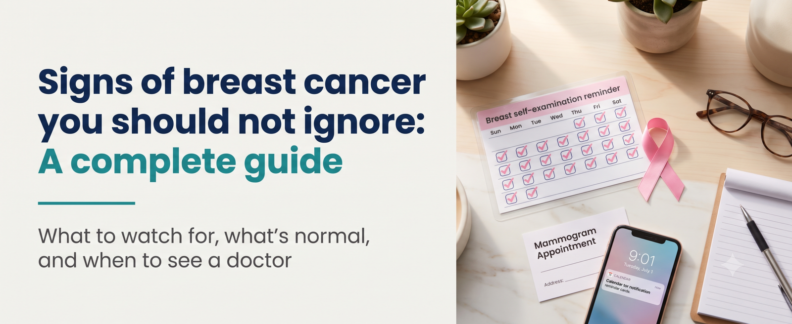
Signs of breast cancer you should not ignore: A complete guide

Signs of breast cancer you should not ignore: A complete guide

What to watch for, what’s normal, and when to see a doctor

This page is for informational purposes only and does not replace medical advice. Please consult Dr Aditya Sarin or a qualified oncologist to evaluate any breast changes specific to you. As of 2026, this reflects current clinical guidance.

Most women’s breasts feel a little lumpy, and that lumpiness on its own usually means nothing. The tricky part is telling the difference between normal texture and something worth checking. Mammograms catch a lot of breast cancers before they cause any symptoms at all, but they don’t catch every case, which is exactly why knowing what your breasts normally look and feel like matters as much as the screening itself.

The most common signs of breast cancer

A new lump or mass is the symptom most people associate with breast cancer, and according to the American Cancer Society, it’s still the most common one. A lump that’s hard, painless, fixed in place, and has irregular edges is more likely to be cancer, though breast cancer can just as easily show up as something soft, round, or tender. 

Beyond a lump, a few other changes are worth paying attention to. Swelling of part or all of the breast, even without a lump you can feel. Skin dimpling that can look like an orange peel. Redness, dryness, flaking, or thickening of the nipple or breast skin. Pain in the breast or nipple. A nipple that turns inward when it didn’t before. Discharge from the nipple that isn’t breast milk. And swollen lymph nodes under the arm or near the collarbone, which can sometimes show up before a lump is even large enough to feel.

None of these alone confirms cancer. What matters is noticing when something is different from your own normal, and getting it checked rather than assuming it’s nothing.

What can cause a breast lump besides cancer?

Most breast lumps aren’t cancerous, and there are several common, harmless reasons a lump can show up, covered in detail in the American Cancer Society’s guide to noncancerous breast conditions. Fibroadenomas are solid, benign lumps that are especially common in younger women. Cysts are fluid-filled sacs that can feel firm and sometimes tender, particularly around your menstrual cycle. Fibrocystic changes cause general lumpiness and tenderness tied to hormone fluctuations, and tend to ease up after menopause. 

Hormones play a big role. Breast tissue changes throughout your menstrual cycle, during pregnancy, and around menopause, and hormonal medications like birth control or hormone replacement therapy can shift how your breasts feel too. None of this means you should ignore a new lump; it just means a benign explanation is more likely, and a doctor can tell the difference with an exam or imaging.

Breast cancer risk factors in India

Breast cancer is the most common cancer among Indian women, accounting for roughly 28.8 percent of cancer cases among women in the country, according to National Cancer Registry Programme estimates published in the Indian Journal of Medical Research. On average, about 1 in 29 Indian women will develop breast cancer during her lifetime, with incidence rising notably from the early thirties and peaking between ages 50 and 64. 

Some risk factors can’t be changed. Family history and inherited mutations in the BRCA1 and BRCA2 genes meaningfully raise risk, as does simply getting older, particularly after menopause. Starting periods early or reaching menopause late also plays a role. Other factors are at least partly modifiable, including obesity, physical inactivity, alcohol use, and long-term use of hormone therapy. A family history of breast, ovarian, or colorectal cancer is a reasonable prompt to talk to a doctor about genetic counselling, even without symptoms yet.

How to do a breast self-examination

Breast self-exams are worth putting in honest context. Major cancer centres, including Memorial Sloan Kettering, no longer recommend BSE as a routine screening tool on its own, since large studies haven’t shown that it reduces breast cancer deaths, and it can sometimes lead to unnecessary worry or biopsies for harmless findings. What actually matters more is breast self-awareness, simply knowing what’s normal for your body so you notice real changes. 

If you’d still like to do a self-exam to build that awareness, here’s a straightforward approach. Stand in front of a mirror with your arms on your hips and look for any changes in size, shape, or skin texture. Raise each arm slightly and check the underarm area for lumps. Then lie down, since breast tissue spreads out and is easier to feel this way, and use the pads of three fingers to check the whole breast in an up-and-down pattern, using light, medium, and firm pressure at each spot. Repeat on the other side using your opposite hand.

Do this once a month if you choose to, ideally a few days after your period ends when breasts are least likely to be tender. But self-exams are a personal choice, not a replacement for a clinical breast exam or mammogram, and skipping them doesn’t put you at any disadvantage as long as you stay aware of your body and get regular screening.

When to see a doctor

Any new lump, persistent pain, skin change, or nipple discharge that isn’t breast milk deserves a proper look from a healthcare professional, even if it seems minor. This is true whether or not you’re currently within your recommended screening age, since symptoms can appear between scheduled mammograms. Getting a change checked early, and confirmed as harmless or caught while still treatable, is always the better outcome than waiting to see if it goes away.

The takeaway

Most breast changes aren’t cancer, but the ones that are get caught earlier and treated more successfully when they’re acted on quickly rather than watched for months. Knowing what’s normal for your own body is more useful day-to-day than any single checklist, and it’s what makes a real change stand out when it happens.

If you’ve noticed a lump, skin change, or anything that feels different,  book a consultation with Dr Aditya Sarin at Sir Ganga Ram Hospital, New Delhi.

References

  1. American Cancer Society: Signs and Symptoms of Breast Cancer.
  2. American Cancer Society: Noncancerous Breast Conditions.
  3. Memorial Sloan Kettering Cancer Centre: Breast Self-Awareness and How To Do a Breast Self-Exam (BSE).
  4. Sathishkumar K, Chaturvedi M, Das P, Stephen S, Mathur P: Cancer incidence estimates for 2022 & projection for 2025: Results from National Cancer Registry Programme, India. Indian J Med Res. 2022;156(4-5):598-607.

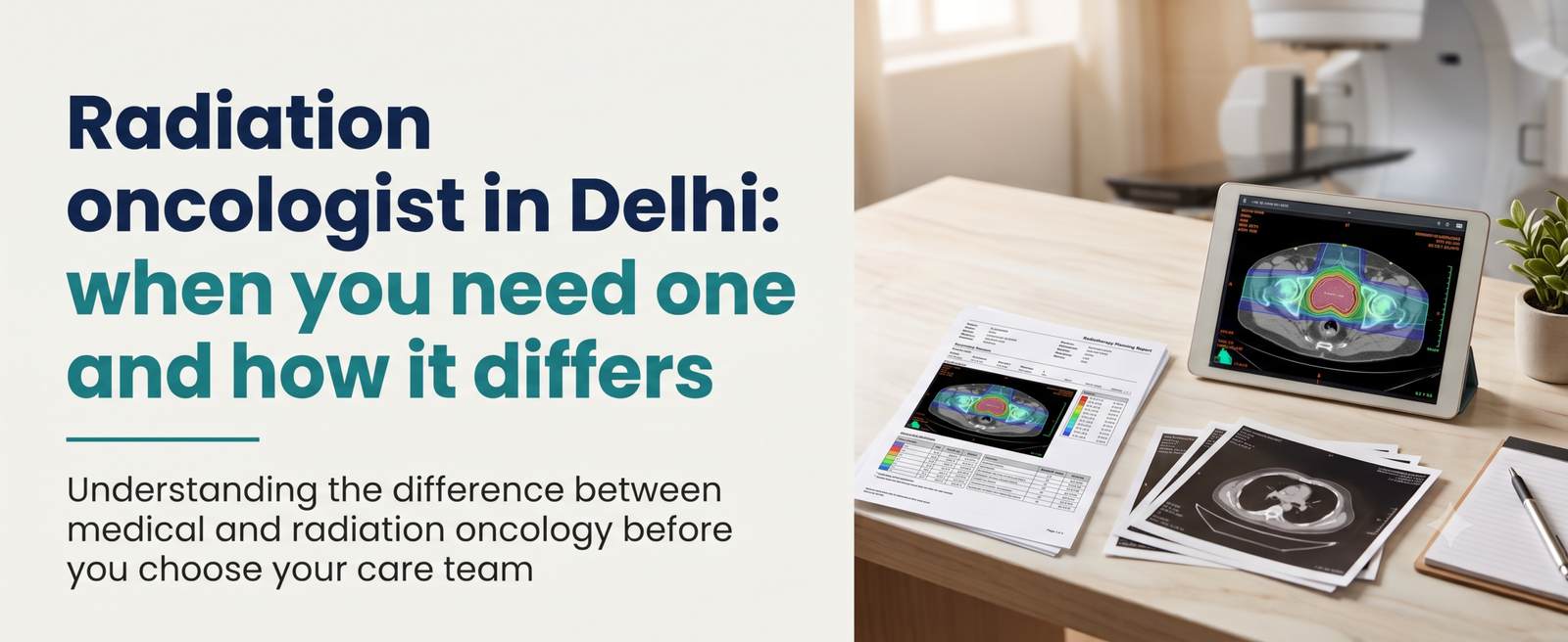
Radiation oncologist in Delhi: when you need one and how it differs

Radiation oncologist in Delhi: when you need one and how it differs

Understanding the difference between medical and radiation oncology before you choose your care team

This page is for informational purposes only and does not replace medical advice. Please consult Dr Aditya Sarin or a qualified oncologist to understand which specialists your specific diagnosis actually requires. As of 2026, this reflects current clinical guidance.

If you or someone close to you has just been diagnosed with cancer, you’ve probably already run into a confusing wall of job titles. Medical oncologist. Radiation oncologist. Surgical oncologist. They all sound similar, but they do genuinely different jobs, and knowing which one you need, and when, can save you a lot of confused phone calls. Here’s what a radiation oncologist actually does, how that differs from a medical oncologist, and how the two work together on an actual treatment plan.

What does a radiation oncologist do

A radiation oncologist is a doctor who specialises in treating cancer using high-energy radiation to destroy cancer cells or shrink tumours. According to the National Cancer Institute, radiation therapy works by damaging the DNA inside cancer cells so they can no longer grow or divide.

Radiation oncologists don’t just point a machine and press a button. They work with a medical physicist and a radiation therapist to plan exactly how much radiation to give, from what angles, and over how many sessions, so the tumour gets the dose it needs while sparing healthy surrounding tissue as much as possible.

What does a medical oncologist do

A medical oncologist treats cancer with medicines that work throughout the body, including chemotherapy, hormone therapy, targeted therapy, and immunotherapy. According to Maryland Oncology Haematology, a medical oncologist often serves as the primary point of contact for a cancer patient, coordinating care and managing side effects across the entire treatment journey, not just one phase.

This is the role Dr Aditya Sarin practices in. His work centres on diagnosing cancer, interpreting biomarker and genetic test results, and building a systemic treatment plan, whether that’s chemotherapy, targeted therapy, or immunotherapy, tailored to a patient’s specific tumour biology.

Medical oncologist vs radiation oncologist: The core difference

The simplest way to think about it: a medical oncologist treats cancer with medicine that travels through the body, while a radiation oncologist treats a specific, defined area with focused radiation. Neither approach is inherently stronger or weaker. They target the disease differently, and which one, or which combination, makes sense depends entirely on the cancer type, its stage, and where it is in the body.

According to Acibadem Hospitals Group, these specialities aren’t in competition with each other. In most real-world cancer care, a patient sees more than one type of oncologist, and the treatment plan is built as a coordinated effort between them, not a choice between two separate paths.

Side effects also differ. Medical oncology treatments can affect the whole body depending on the drug involved, while radiation side effects are usually limited to the treated area, though they still vary depending on where that area is.

When do you actually need a radiation oncologist

Not every cancer diagnosis calls for radiation, and that’s an important thing to understand upfront rather than assuming every patient’s team looks the same. Radiation is generally considered when one or more of the following applies.

The cancer is confined to a specific, localised area, which makes it a good candidate for focused treatment rather than a whole-body approach.

Surgery has removed a visible tumour, but there’s a meaningful risk of microscopic cancer cells remaining nearby, in which case radiation is used afterwards to reduce the chance of recurrence.

The tumour needs to be shrunk before surgery to make the operation more feasible or less extensive.

The goal is symptom relief rather than cure, such as easing pain from a tumour that has spread to bone, where radiation can meaningfully improve quality of life even in advanced disease.

Cancers of the head and neck, breast, cervix, prostate, and certain blood cancers like lymphoma commonly involve radiation as part of the overall plan, according to the National Cancer Institute, though the exact role varies by case.

The main types of radiation therapy

Radiation therapy isn’t one single technique. The two broad categories are external beam radiation therapy, delivered from a machine outside the body and the most widely used approach, and brachytherapy, sometimes called internal radiation, where a radioactive source is placed directly inside or next to the tumour. Brachytherapy is used most often for cancers of the head and neck, breast, cervix, and prostate.

There are also systemic forms of radiation therapy, such as radioactive iodine for certain thyroid cancers, that work more like a targeted internal treatment than a localised beam.

For most people, radiation isn’t a stand-alone treatment. It’s typically combined with surgery, chemotherapy, or immunotherapy, and a medical oncologist helps coordinate that across the full course of care.

How a medical oncologist and radiation oncologist work together

In practice, cancer care rarely comes down to one specialist working in isolation. A patient with breast cancer, for example, might see a surgeon to remove the tumour, a radiation oncologist for follow-up radiation to the chest wall, and a medical oncologist managing hormone therapy or chemotherapy over the following months, all as part of one coordinated plan. The same layered approach applies to cervical cancer, where chemotherapy and radiation are frequently used together, and to several throat cancer cases, where radiation, surgery, and systemic therapy are often combined.

According to ASCO Connection, the American Society of Clinical Oncology’s own platform, modern cancer care runs on multidisciplinary teams where medical oncologists, radiation oncologists, and other specialists each bring a different role to a patient’s case. Rather than patients choosing between them, a coordinated, jointly reviewed treatment plan is generally considered the standard of care. 

Where does a medical oncologist fit into this decision?

If you’re newly diagnosed and unsure who to see first, a medical oncologist is often the right starting point. Because the role involves coordinating the broader treatment plan and interpreting diagnostic and biomarker results, a medical oncologist can help determine whether radiation, chemotherapy, targeted therapy, surgery, or some combination is the right path, and refer you to the appropriate specialists accordingly, rather than you needing to guess which type of oncologist to book first.

A second opinion can also be genuinely useful, particularly if you’ve already been told you need radiation and want an independent review of whether that’s the most appropriate next step given your specific diagnosis and stage.

The takeaway

Medical and radiation oncology aren’t competing paths; they’re complementary parts of modern cancer care, and most patients benefit from both being involved at the right point in treatment. If you’ve been diagnosed and aren’t sure which specialists your case actually needs, book a consultation with Dr Aditya Sarin to get a clear, coordinated starting point for your treatment plan.

References

  1. National Cancer Institute: Radiation Therapy for Cancer
  2. Maryland Oncology Haematology: Understanding Your Cancer Care Team
  3. ASCO Connection, American Society of Clinical Oncology: Contemporary Clinical Oncology Training in the United States: Is Radiation or Medical Oncology Right for You?.

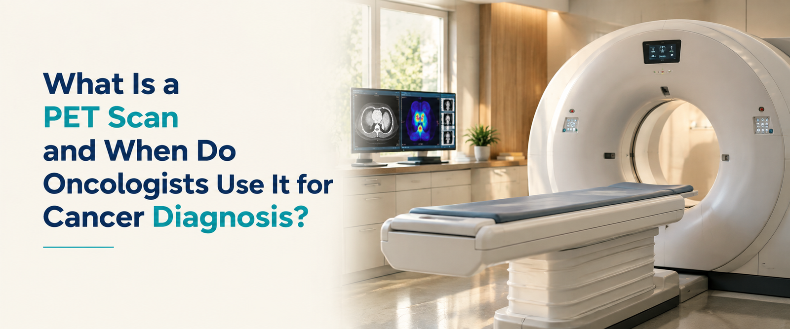
What Is a PET Scan? Uses, Cost and Results Explained

What is a PET scan and when do oncologists use it for cancer diagnosis?

A plain-language guide to how PET scans work, how they differ from a CT scan, what results actually mean, and what they cost in India.

Summary

A PET scan is an imaging test that shows how active tissue is at a cellular level, rather than just what it looks like structurally. Oncologists use it mainly for staging a new cancer diagnosis, checking whether treatment is working, and looking for recurrence after treatment ends. This guide covers how the scan works, how it differs from a CT scan, how to read your results, and what it typically costs in India.

This content is for informational purposes only and does not constitute medical advice. Your treating oncologist should decide whether a PET scan is appropriate for your situation. As of 2026.

If you’ve been told you need a PET scan, or a PET-CT specifically, it can feel like one more unfamiliar step in an already overwhelming process. The name itself doesn’t help. Positron emission tomography sounds intimidating, but the underlying idea is actually fairly intuitive once it’s explained properly.

This guide walks through exactly that.

What is a PET scan?

A PET scan is an imaging test that shows how metabolically active different tissues in the body are by tracking where a small amount of radioactive tracer, most commonly a glucose analogue called FDG, gets taken up in the body.

The core idea behind it is simple. Cancer cells generally grow and divide faster than normal cells, and that growth takes energy. Fast-growing cells tend to consume far more glucose than normal tissue does. When a patient is injected with FDG, a radioactive form of glucose, cells that are working harder, including most cancer cells, absorb more of it. The PET scanner then detects where that tracer has concentrated and builds an image showing those areas of high activity.

This is fundamentally different from most other imaging. A CT or MRI shows what a structure looks like. A PET scan shows what it’s actually doing.

How does a PET scan actually work?

The process itself is fairly straightforward for the patient, even though the science behind it is sophisticated.

You’re given an injection of the FDG tracer, usually into a vein in the arm. Then there’s a waiting period, typically around 60 minutes, while the tracer circulates through the body and is taken up by active tissue. During this time, you’re usually asked to rest quietly, since muscle activity or talking can cause the tracer to accumulate in places unrelated to any tumour, muddying the results. After the waiting period, you lie on a scanning table that moves slowly through the PET scanner, which typically takes 20 to 45 minutes depending on how much of the body is being imaged.

When do oncologists actually order a PET scan?

PET scans come up at several distinct points in cancer care, and it helps to know which one applies to your situation.

Initial staging: Once a cancer diagnosis is confirmed, a PET scan helps determine how far it has spread, to nearby lymph nodes or to distant organs, which directly shapes the treatment plan. In several cancers, including lung cancer, PET has been shown to be more sensitive than CT alone for detecting nodal involvement and distant spread, and can change the assigned stage and recommended treatment in a meaningful share of patients. This kind of imaging often works alongside the molecular and genomic testing that increasingly guides personalised treatment decisions.

Treatment response assessment: A PET scan partway through treatment can show whether a tumour’s metabolic activity is dropping in response to chemotherapy or radiation, sometimes well before any change would be visible on a structural scan like CT.

Detecting recurrence: After treatment ends, a PET scan can help investigate whether cancer has returned, particularly when blood markers or symptoms raise suspicion but a standard CT scan isn’t conclusive.

Radiation treatment planning: In several cancers, PET imaging helps radiation oncologists define exactly which tissue to target, since it distinguishes active tumour from surrounding scar tissue or collapsed lung more precisely than CT alone.

Not every cancer diagnosis requires a PET scan, and not every stage of treatment calls for one. Your oncologist will recommend it specifically when the information it provides is likely to change a decision.

What is the difference between a PET scan and a CT scan?

This is one of the most common points of confusion, and the distinction matters practically.

Factor

CT scan

PET scan

What it shows

Anatomical structure, size, shape, and location

Metabolic activity, how active the tissue is

Best at

Detailed anatomy, precise measurements, guiding biopsies

Detecting abnormal activity, even in normal-looking tissue

Typical use

Structural detail, surgical planning, routine follow-up imaging

Staging, treatment response, detecting recurrence

Radiation source

X-rays

A small dose of radioactive tracer, plus a low-dose CT in most combined scans

In practice, oncologists rarely choose one over the other in isolation. They’re usually looking for both pieces of information at once, which is why combined PET-CT has become the standard approach in most cancer centres.

What is a PET-CT scan, and why are they usually done together?

A PET-CT scan combines both technologies in a single machine and a single appointment. The CT portion provides the detailed anatomical map, while the PET portion overlays exactly where the metabolic activity is happening on that map.

The combination solves a real limitation of PET on its own. A PET scan can show a bright spot of activity, but without precise anatomical detail, it can be hard to tell exactly which structure that spot corresponds to. Fusing it with CT imaging pinpoints the finding to an exact location, which is far more clinically useful than either scan alone.

How are PET scan results explained?

PET scan reports usually include a number called the standardised uptake value, or SUV, alongside the images themselves. It’s worth understanding roughly what this number means, since it comes up in almost every PET report.

SUV is a ratio that quantifies how much tracer has accumulated in a specific area, adjusted for the dose given and the patient’s body weight. A higher SUV generally means higher metabolic activity in that spot. As a rough reference point, healthy blood typically shows an SUV between 1.5 and 1.9, and the liver usually sits around 2.3 to 2.8, so radiologists compare suspicious areas against these normal background levels rather than reading a number in isolation. 

Here’s the detail that matters most for interpreting your own results. A high SUV is not automatically cancer. Inflammation, infection, and even normal muscle activity from talking or fidgeting during the uptake period can raise SUV in a specific area. What your radiologist and oncologist are actually looking at is the pattern, the specific location, the shape of the uptake, and how it compares with your prior scans if you have any, not a single number taken in isolation. If you’re monitoring treatment response, the trend across scans over time tends to matter more than any single value.

What does a PET scan cost in India?

Cost varies considerably depending on the facility type and the specific scan ordered.

Scan type

Typical cost range in India (2026)

Whole-body FDG PET-CT, standalone diagnostic centre

Roughly ₹8,000 to ₹20,000

Whole-body FDG PET-CT, large private hospital

Roughly ₹25,000 to ₹40,000

PSMA PET-CT (used for prostate cancer)

Roughly ₹17,500 to ₹30,000

Dotanoc or DOTA PET-CT (used for neuroendocrine tumours)

Roughly ₹18,000 to ₹28,000

These are approximate ranges based on current Delhi NCR pricing data; actual pricing depends on the city, the specific centre, and whether the tracer and radiologist’s report are included in the quoted price. It’s worth asking directly whether a quote covers the tracer dose, the scan itself, the nuclear medicine physician’s report, and image delivery, since additional charges tacked on afterwards are a common source of unexpected bills. Government hospitals often offer meaningfully subsidised rates for eligible patients, which is worth asking about directly if cost is a concern.

What are the limitations of a PET scan?

It’s worth being upfront about this rather than presenting PET as a perfect tool. Very small tumours, generally under about a centimetre, can be missed, since there isn’t enough tissue mass to produce a clearly detectable signal. Some slow-growing cancers don’t take up much glucose at all, which can make them harder to detect on FDG-PET specifically, regardless of tumour size. And as covered above, a positive-looking area on a PET scan doesn’t automatically confirm cancer, since inflammation and infection can produce similar signals.

None of this makes PET scans unreliable. It means a radiologist and your treating oncologist interpret results together, in the context of your specific history, other test results, and, where needed, a biopsy to confirm what the scan is actually showing. 

How should you prepare for a PET scan?

Preparation is fairly consistent across most centres, though it’s always worth confirming specific instructions with the facility performing your scan.

You’ll typically be asked to fast for four to six hours beforehand, since eating raises blood glucose levels, which can interfere with how the tracer distributes in the body. Diabetic patients often need specific guidance on managing blood sugar and medication timing before the scan, so this is worth discussing with your doctor in advance. You’ll usually be asked to avoid strenuous exercise for a day or two beforehand, since muscle activity can affect uptake patterns, and to wear comfortable clothing without metal, since it can interfere with the CT portion of a combined scan.

The takeaway

A PET scan gives oncologists a genuinely different kind of information than a standard CT or MRI, showing how active tissue is rather than just what it looks like. That makes it a valuable tool at several distinct points in cancer care, from initial staging through to checking whether treatment is working. Understanding what the results mean, and what reasonable questions to ask, makes the process considerably less daunting.

Book a consultation with Dr Aditya Sarin at Sir Ganga Ram Hospital, New Delhi

Colon cancer symptoms, screening and what to do next

Colon cancer symptoms, screening and what to do next

A clear, practical guide to the warning signs, when to get screened, and what actually happens if something is found.

Summary

Colon cancer, part of the broader group called colorectal cancer, often develops silently for years before it causes any obvious symptoms. This guide covers the warning signs worth taking seriously, why the disease is rising sharply in younger adults, when screening should actually start, and what practical steps to take next if you’re worried or if a screening test comes back abnormal.

This content is for informational purposes only and does not constitute medical advice. Many of the symptoms described here have common, non-cancerous causes. Always consult a qualified doctor for evaluation, diagnosis, and treatment specific to your situation. As of 2026.

Colon cancer has a reputation as an older person’s disease, and for a long time that was largely true. That’s changing quickly, and not in a good way. Rates in adults under 50 have been climbing for three decades, and colorectal cancer is now the leading cause of cancer death in men under 50 in the United States, and the second leading cause in women under 50. The reasons for that shift aren’t fully understood yet, but the practical takeaway is simple. This isn’t a disease you can safely assume you’re too young for.

This guide is built to help you recognise what actually matters, and know what to do about it.

What are the early symptoms of colon cancer?

Early colon cancer often causes no symptoms at all, which is exactly why screening matters so much. When symptoms do appear, a handful come up again and again.

  • A change in bowel habits, diarrhoea, constipation, or narrower stools that lasts more than a few days.
  • Blood in the stool, or on toilet paper, ranging from bright red to dark and tarry.
  • A feeling that your bowel hasn’t fully emptied, even right after a bowel movement.
  • Ongoing abdominal pain, cramping, or bloating that doesn’t resolve.
  • Unexplained fatigue or weakness, which can come from anaemia caused by slow, hidden blood loss.
  • Unintentional weight loss.

None of these symptoms is unique to colon cancer. Each one of them is far more commonly caused by something else entirely: haemorrhoids, irritable bowel syndrome, an infection, or a dietary change. That’s what makes them easy to dismiss, and it’s also why persistence matters most. A symptom that shows up once and disappears is different from one that lingers for weeks. The American Cancer Society’s full symptom list is a useful reference if you want to check something specific.

Why do so many people miss these symptoms?

Partly because the symptoms overlap so heavily with common, harmless conditions, and partly because talking about bowel habits still makes people uncomfortable enough to put off a conversation they’d have quickly about almost anything else.

There’s a second, more specific reason worth naming directly. Unexplained iron-deficiency anaemia, showing up as fatigue, weakness, or shortness of breath on a routine blood test, is one of the more overlooked warning signs. It doesn’t announce itself as a digestive symptom, and it’s sometimes chalked up to diet or a busy schedule rather than investigated further. If a blood test shows unexplained anaemia and it doesn’t resolve with an obvious explanation, that’s a reasonable moment to ask directly whether a colonoscopy is warranted.

What causes colon cancer?

Most colon cancer starts as a polyp, a small growth on the inner lining of the colon that is not initially cancerous. Over years, some polyps accumulate genetic changes that allow them to become cancerous. This slow progression is actually good news, because it creates a genuine window for screening to catch and remove polyps before they ever turn into cancer.

Several factors raise the likelihood of this happening.

  • Age, with risk rising steadily after 45, though it’s increasingly showing up younger.
  • A personal or family history of colorectal polyps or cancer.
  • Inflammatory bowel disease, such as ulcerative colitis or Crohn’s disease.
  • Inherited conditions such as Lynch syndrome or familial adenomatous polyposis.
  • Diets low in fibre and high in red or processed meat.
  • Obesity, physical inactivity, smoking, and heavy alcohol use.
  • Type 2 diabetes.

Having one or more of these factors doesn’t guarantee colon cancer will develop, and plenty of people diagnosed have no clear risk factor beyond age. But several of these, diet, activity level, smoking, and alcohol, are genuinely modifiable, which is part of why colon cancer is considered one of the more preventable cancers overall.

Why is colon cancer rising in younger people, in India and everywhere else?

This trend is real, global, and still not fully explained. In the United States, about 1 in 5 colorectal cancer cases is now diagnosed in someone under 54, compared with roughly 1 in 10 three decades ago. Researchers suspect diet, physical inactivity, obesity, and other environmental exposures play a role, but no single cause has been confirmed.

India shows a similar pattern, with a few distinct features worth knowing. Colorectal cancer is now the fourth most common cancer among men in India, and incidence has been climbing across nearly every Indian cancer registry, according to a review in the Indian Journal of Cancer. The average age at diagnosis in India tends to be younger than in Western countries, with some tertiary centre studies reporting a mean age around 47, and up to a third of cases occurring before age 40. Indian studies specifically looking at young-onset colorectal cancer have found these cases are more likely to present at an advanced stage and more likely to involve an aggressive histological subtype called signet-ring cell carcinoma.

The presentation pattern in India is also skewed later than ideal. In one Indian dataset, only about 4% of cases were caught at Stage I, while over a quarter were already Stage IV at diagnosis. That gap between what’s possible with early detection and what’s actually happening on the ground is really the central problem this guide is trying to address.

When should you start colon cancer screening?

Situation

Recommended approach

Average risk, United States guidelines (ACS, USPSTF)

Begin screening at age 45 and continue through 75.

Average risk, India

No formal national screening guideline currently exists. Experts increasingly recommend discussing screening with a doctor from around 45, especially in urban settings with rising incidence.

Family history of colorectal cancer or polyps

Often recommended to start earlier, sometimes 10 years before the age the relative was diagnosed. Discuss timing directly with your doctor.

Inflammatory bowel disease or a known genetic syndrome

Screening typically starts earlier and happens more frequently. Plan this with a specialist.

It’s worth being direct about something here. India doesn’t yet have a unified, national colorectal cancer screening guideline the way the United States does. Several Indian medical bodies have called for one given the rising incidence, but until that exists, the practical approach is a personal conversation with a doctor, particularly from your mid-40s onward, or earlier with any family history or persistent symptoms.

What are the screening tests available?

Screening broadly falls into two categories: tests that look directly at the colon, and tests that check a stool sample for hidden signs of blood or abnormal cells.

Colonoscopy is the most thorough option, allowing a doctor to examine the entire colon and remove polyps in the same procedure, before they ever have the chance to become cancerous. It’s typically repeated every 10 years for average-risk people with a normal result.

Faecal immunochemical testing (FIT) is a simple, non-invasive stool test that checks for hidden blood, done annually. It has a reported sensitivity around 97% and a negative predictive value near 99.8%, making it a genuinely useful, low-cost triage tool, particularly relevant in settings where colonoscopy access is limited.

Flexible sigmoidoscopy examines the lower part of the colon and is sometimes used alongside stool-based testing, though it’s less commonly used as a standalone option than it once was.

CT colonography, a specialised imaging scan, is an alternative for people who can’t or prefer not to have a standard colonoscopy.

No single test is automatically the right choice for everyone. The right one depends on personal risk, access, and what a doctor recommends based on your specific situation. 

Our guide on cancer diagnostics and early detection covers how these tests fit into the bigger picture of early cancer detection.

What happens if something is found.

If a screening test finds a polyp or something abnormal, the next step is almost always a colonoscopy, if one hasn’t already been done, since it allows both diagnosis and, for most polyps, immediate removal in the same procedure.

If a biopsy confirms cancer, the next step is staging, determining how far, if at all, the cancer has spread beyond the colon wall. This most directly shapes the treatment plan and outlook. The difference staging makes is substantial. Localised colon cancer, caught before it has spread beyond the colon, has a 5-year relative survival rate of around 90%, according to NCI SEER data. Once it has spread to distant organs, that figure drops to roughly 15%. That gap is really the entire argument for screening in one statistic. 

If you’ve already received a diagnosis and want a second set of eyes on the plan, our second opinion page explains how that process works.

How is colon cancer treated?

Treatment depends heavily on the stage at diagnosis, and it’s rarely just one approach.

Surgery to remove the cancerous section of the colon is the primary treatment for most non-metastatic colon cancer, and is often curative on its own for early-stage disease.

Chemotherapy is commonly added after surgery for higher-risk or more advanced stages, to reduce the chance of recurrence, and is central to treatment once the cancer has spread. Our chemotherapy page explains how this works in more detail.

Targeted therapy and immunotherapy are used for select advanced or metastatic cases, often guided by molecular testing of the tumour to identify which specific drugs are likely to work. See our pages on targeted therapy and immunotherapy for more on how each approach is used.

Radiation therapy plays a larger role in rectal cancer specifically than in colon cancer higher up the digestive tract, often combined with chemotherapy before surgery.

The right combination depends entirely on stage, tumour location, and, increasingly, the specific molecular profile of the tumour, which is why an individualised treatment plan matters more than a generic protocol.

What should you actually do next?

If you have a persistent symptom from the list above, don’t wait it out hoping it resolves on its own. See a doctor, describe exactly what’s happening and for how long, and let them guide the next step, whether that’s further testing or simple reassurance.

If you’re over 45, or have a family history, and haven’t had a screening conversation with a doctor yet, that conversation itself is the next step, not a full colonoscopy on day one. A doctor can help you weigh options, including FIT testing as a lower-barrier starting point.

If a screening test has already come back abnormal, follow through with the recommended next test rather than sitting on the result. The entire value of screening depends on acting on what it finds.

The takeaway
Colon cancer is genuinely one of the more preventable and treatable cancers, but only when it’s caught before it has had time to spread. Persistent symptoms deserve a proper look rather than an assumption, and given how much earlier this disease is now showing up, screening conversations shouldn’t wait for a specific trigger.
Book a consultation with Dr Aditya Sarin at Sir Ganga Ram Hospital, New Delhi


Tumour markers explained: What they tell your doctor about cancer

A plain-language guide to CA 125, CEA, and the blood tests behind your cancer care

This page is for informational purposes only and does not replace medical advice. Please consult Dr Aditya Sarin or a qualified oncologist to understand what your specific tumour marker results mean. As of 2026, this reflects current clinical guidance.

If you’ve had bloodwork done as part of a cancer diagnosis, you’ve probably seen a result labelled CA 125 or CEA and had no idea what it actually meant. Tumour markers show up on a lot of lab reports, but they’re rarely explained well. Here’s what they are, what the common ones actually track, and where they fit into an actual treatment plan.


What is a tumour marker?

A tumour marker is a substance, usually a protein, found in blood, urine, or tissue that can be elevated when cancer is present. According to the National Cancer Institute, tumour markers are mainly used to help diagnose cancer, guide treatment decisions, and monitor how well a treatment is working, not to screen healthy people for cancer in most cases.

That last part matters. Tumour markers aren’t a stand-alone way to find cancer early. With the exception of PSA for prostate cancer, most markers don’t have the accuracy needed for population screening. They’re better understood as one piece of a larger picture that includes imaging, biopsy, and clinical evaluation.


What are the main types of tumour markers?

There are dozens of tumour markers in clinical use, but a handful come up constantly. Each is associated with certain cancer types, though none is exclusive to just one.

CA 125 is most closely linked to ovarian cancer. It’s elevated in roughly 80 per cent of ovarian cancer cases, but it also rises with completely unrelated things like endometriosis, pregnancy, and menstruation, so a high result alone doesn’t confirm cancer.

CEA, or carcinoembryonic antigen, is used primarily to monitor colorectal cancer, both to track treatment response and to catch recurrence after surgery. It’s not used to diagnose colon cancer in the first place, since plenty of non-cancerous conditions, including smoking, can raise it too.

CA 19-9 is associated mainly with pancreatic cancer and is also used in some gastrointestinal cancers. It helps distinguish the nature of a pancreatic mass and track how treatment is going.

AFP, or alpha-fetoprotein, is the main marker for liver cancer and is also used for certain testicular cancers.

PSA is specific to prostate cancer and is the one marker sensitive enough to be used in actual screening programs, though even PSA has known limitations around false positives.

CA 15-3 and CA 27-29 are used mainly to monitor treatment response in metastatic breast cancer, not for initial diagnosis.


What does a tumour marker blood test actually show

A tumour marker test measures the concentration of that specific substance in your blood at one point in time. On its own, a single number rarely tells the full story. What matters more is the trend.

If CA 125 or CEA is rising steadily over several tests, that’s a stronger signal than one high reading. If a marker drops after surgery or chemotherapy and stays low, that generally means treatment is working. If it starts climbing again after being low, that can be an early sign of recurrence, sometimes months before it would show up on a scan.

This is why oncologists tend to order these tests repeatedly through a treatment course rather than once. The pattern over time carries more information than any single result.


Can a tumour marker diagnose cancer on its own

No. This is probably the most important thing to understand about tumour markers. According to the American Cancer Society, an elevated marker can point toward cancer, but confirming a diagnosis still requires imaging and, in most cases, a biopsy.

Several conditions that have nothing to do with cancer can raise these same markers. CA 125 rises with pelvic inflammatory disease. CEA rises in smokers and with inflammatory bowel disease. A normal marker level also doesn’t rule out cancer entirely, since not every tumour produces measurable amounts of a given marker. Some ovarian cancers, for instance, never raise CA 125 at all.


Tumour marker testing in India

Tumour marker blood tests are widely available across major diagnostic labs in India and are a standard part of oncology workups at hospitals like Sir Ganga Ram Hospital. They’re typically ordered alongside imaging such as CT or PET scans rather than in isolation, which is the approach Dr Aditya Sarin follows when building a diagnostic picture for a patient. For markers tied to specific cancers, CA 125 for ovarian cancer, CEA for colorectal cancer, CA 19-9 for pancreatic cancer, or AFP for liver cancer, testing is usually combined with biomarker and genetic profiling to guide not just diagnosis but the choice between treatments like chemotherapy, targeted therapy, or immunotherapy.


The takeaway

Tumour markers are a useful piece of the puzzle, not the whole picture. They’re best at tracking trends over time and monitoring how well treatment is working, and least reliable when used alone to diagnose or rule out cancer. If you’ve got a tumour marker result you don’t understand, book a consultation to have it explained in the context of your actual case.