
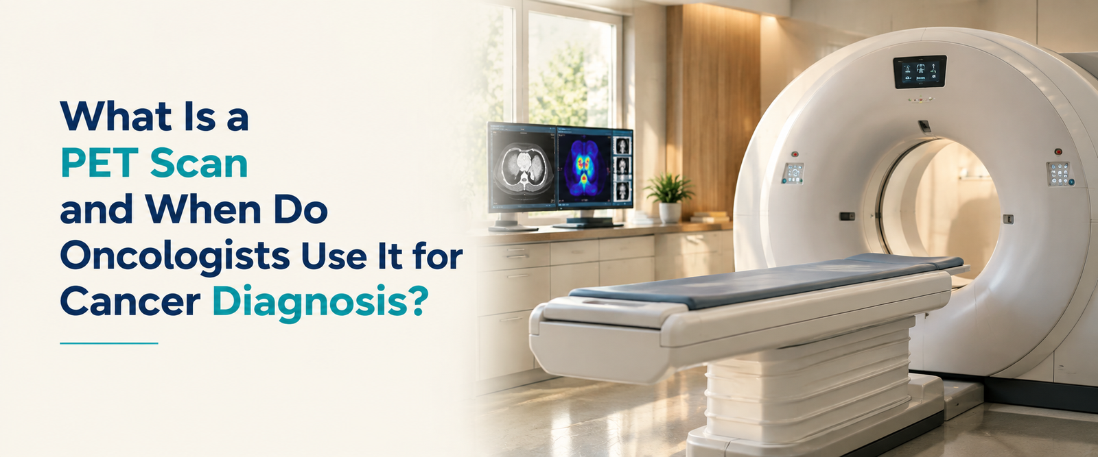
What is a PET scan and when do oncologists use it for cancer diagnosis?
A plain-language guide to how PET scans work, how they differ from a CT scan, what results actually mean, and what they cost in India.
Summary
A PET scan is an imaging test that shows how active tissue is at a cellular level, rather than just what it looks like structurally. Oncologists use it mainly for staging a new cancer diagnosis, checking whether treatment is working, and looking for recurrence after treatment ends. This guide covers how the scan works, how it differs from a CT scan, how to read your results, and what it typically costs in India.
This content is for informational purposes only and does not constitute medical advice. Your treating oncologist should decide whether a PET scan is appropriate for your situation. As of 2026.
If you’ve been told you need a PET scan, or a PET-CT specifically, it can feel like one more unfamiliar step in an already overwhelming process. The name itself doesn’t help. Positron emission tomography sounds intimidating, but the underlying idea is actually fairly intuitive once it’s explained properly.
This guide walks through exactly that.
What is a PET scan?
A PET scan is an imaging test that shows how metabolically active different tissues in the body are by tracking where a small amount of radioactive tracer, most commonly a glucose analogue called FDG, gets taken up in the body.
The core idea behind it is simple. Cancer cells generally grow and divide faster than normal cells, and that growth takes energy. Fast-growing cells tend to consume far more glucose than normal tissue does. When a patient is injected with FDG, a radioactive form of glucose, cells that are working harder, including most cancer cells, absorb more of it. The PET scanner then detects where that tracer has concentrated and builds an image showing those areas of high activity.
This is fundamentally different from most other imaging. A CT or MRI shows what a structure looks like. A PET scan shows what it’s actually doing.
How does a PET scan actually work?
The process itself is fairly straightforward for the patient, even though the science behind it is sophisticated.
You’re given an injection of the FDG tracer, usually into a vein in the arm. Then there’s a waiting period, typically around 60 minutes, while the tracer circulates through the body and is taken up by active tissue. During this time, you’re usually asked to rest quietly, since muscle activity or talking can cause the tracer to accumulate in places unrelated to any tumour, muddying the results. After the waiting period, you lie on a scanning table that moves slowly through the PET scanner, which typically takes 20 to 45 minutes depending on how much of the body is being imaged.
When do oncologists actually order a PET scan?
PET scans come up at several distinct points in cancer care, and it helps to know which one applies to your situation.
Initial staging: Once a cancer diagnosis is confirmed, a PET scan helps determine how far it has spread, to nearby lymph nodes or to distant organs, which directly shapes the treatment plan. In several cancers, including lung cancer, PET has been shown to be more sensitive than CT alone for detecting nodal involvement and distant spread, and can change the assigned stage and recommended treatment in a meaningful share of patients. This kind of imaging often works alongside the molecular and genomic testing that increasingly guides personalised treatment decisions.
Treatment response assessment: A PET scan partway through treatment can show whether a tumour’s metabolic activity is dropping in response to chemotherapy or radiation, sometimes well before any change would be visible on a structural scan like CT.
Detecting recurrence: After treatment ends, a PET scan can help investigate whether cancer has returned, particularly when blood markers or symptoms raise suspicion but a standard CT scan isn’t conclusive.
Radiation treatment planning: In several cancers, PET imaging helps radiation oncologists define exactly which tissue to target, since it distinguishes active tumour from surrounding scar tissue or collapsed lung more precisely than CT alone.
Not every cancer diagnosis requires a PET scan, and not every stage of treatment calls for one. Your oncologist will recommend it specifically when the information it provides is likely to change a decision.
What is the difference between a PET scan and a CT scan?
This is one of the most common points of confusion, and the distinction matters practically.
Factor | CT scan | PET scan |
What it shows | Anatomical structure, size, shape, and location | Metabolic activity, how active the tissue is |
Best at | Detailed anatomy, precise measurements, guiding biopsies | Detecting abnormal activity, even in normal-looking tissue |
Typical use | Structural detail, surgical planning, routine follow-up imaging | Staging, treatment response, detecting recurrence |
Radiation source | X-rays | A small dose of radioactive tracer, plus a low-dose CT in most combined scans |
In practice, oncologists rarely choose one over the other in isolation. They’re usually looking for both pieces of information at once, which is why combined PET-CT has become the standard approach in most cancer centres.
What is a PET-CT scan, and why are they usually done together?
A PET-CT scan combines both technologies in a single machine and a single appointment. The CT portion provides the detailed anatomical map, while the PET portion overlays exactly where the metabolic activity is happening on that map.
The combination solves a real limitation of PET on its own. A PET scan can show a bright spot of activity, but without precise anatomical detail, it can be hard to tell exactly which structure that spot corresponds to. Fusing it with CT imaging pinpoints the finding to an exact location, which is far more clinically useful than either scan alone.
How are PET scan results explained?
PET scan reports usually include a number called the standardised uptake value, or SUV, alongside the images themselves. It’s worth understanding roughly what this number means, since it comes up in almost every PET report.
SUV is a ratio that quantifies how much tracer has accumulated in a specific area, adjusted for the dose given and the patient’s body weight. A higher SUV generally means higher metabolic activity in that spot. As a rough reference point, healthy blood typically shows an SUV between 1.5 and 1.9, and the liver usually sits around 2.3 to 2.8, so radiologists compare suspicious areas against these normal background levels rather than reading a number in isolation.
Here’s the detail that matters most for interpreting your own results. A high SUV is not automatically cancer. Inflammation, infection, and even normal muscle activity from talking or fidgeting during the uptake period can raise SUV in a specific area. What your radiologist and oncologist are actually looking at is the pattern, the specific location, the shape of the uptake, and how it compares with your prior scans if you have any, not a single number taken in isolation. If you’re monitoring treatment response, the trend across scans over time tends to matter more than any single value.
What does a PET scan cost in India?
Cost varies considerably depending on the facility type and the specific scan ordered.
Scan type | Typical cost range in India (2026) |
Whole-body FDG PET-CT, standalone diagnostic centre | Roughly ₹8,000 to ₹20,000 |
Whole-body FDG PET-CT, large private hospital | Roughly ₹25,000 to ₹40,000 |
PSMA PET-CT (used for prostate cancer) | Roughly ₹17,500 to ₹30,000 |
Dotanoc or DOTA PET-CT (used for neuroendocrine tumours) | Roughly ₹18,000 to ₹28,000 |
These are approximate ranges based on current Delhi NCR pricing data; actual pricing depends on the city, the specific centre, and whether the tracer and radiologist’s report are included in the quoted price. It’s worth asking directly whether a quote covers the tracer dose, the scan itself, the nuclear medicine physician’s report, and image delivery, since additional charges tacked on afterwards are a common source of unexpected bills. Government hospitals often offer meaningfully subsidised rates for eligible patients, which is worth asking about directly if cost is a concern.
What are the limitations of a PET scan?
It’s worth being upfront about this rather than presenting PET as a perfect tool. Very small tumours, generally under about a centimetre, can be missed, since there isn’t enough tissue mass to produce a clearly detectable signal. Some slow-growing cancers don’t take up much glucose at all, which can make them harder to detect on FDG-PET specifically, regardless of tumour size. And as covered above, a positive-looking area on a PET scan doesn’t automatically confirm cancer, since inflammation and infection can produce similar signals.
None of this makes PET scans unreliable. It means a radiologist and your treating oncologist interpret results together, in the context of your specific history, other test results, and, where needed, a biopsy to confirm what the scan is actually showing.
How should you prepare for a PET scan?
Preparation is fairly consistent across most centres, though it’s always worth confirming specific instructions with the facility performing your scan.
You’ll typically be asked to fast for four to six hours beforehand, since eating raises blood glucose levels, which can interfere with how the tracer distributes in the body. Diabetic patients often need specific guidance on managing blood sugar and medication timing before the scan, so this is worth discussing with your doctor in advance. You’ll usually be asked to avoid strenuous exercise for a day or two beforehand, since muscle activity can affect uptake patterns, and to wear comfortable clothing without metal, since it can interfere with the CT portion of a combined scan.
The takeaway
A PET scan gives oncologists a genuinely different kind of information than a standard CT or MRI, showing how active tissue is rather than just what it looks like. That makes it a valuable tool at several distinct points in cancer care, from initial staging through to checking whether treatment is working. Understanding what the results mean, and what reasonable questions to ask, makes the process considerably less daunting.
Book a consultation with Dr Aditya Sarin at Sir Ganga Ram Hospital, New Delhi